The eye is a light-sensitive organ and a visual organ. The simplest eyes in the animal kingdom can only distinguish the presence or absence of light. Shapes and colours can be distinguished by the relatively complex structure of the eyes of advanced creatures. Many creatures (including humans) have two eyes on the same floor and form a single three-dimensional "scene". Again, the eyes of many creatures are located on two different surfaces and form two separate scenes (e.g. ; Rabbit eyes)
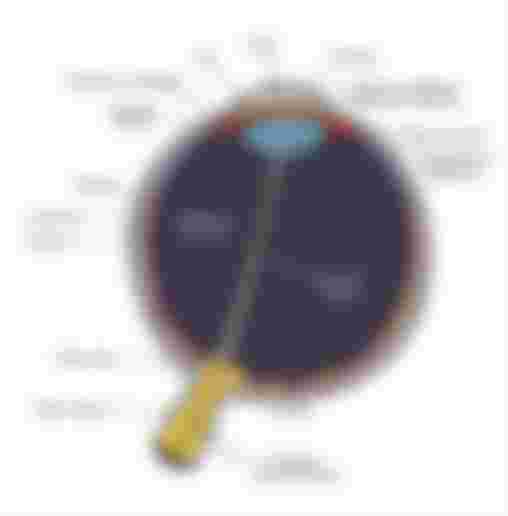
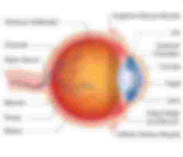
Different parts of the human eye:
Sclera:
It is the white part covering the eyes. It is located 5/6 of the posterior part of the peripheral eye. It and the internal fluids (aquas humour and vitreous humour) together protect the delicate parts of the eye. It is made up of white and opaque and white colored collagen fibers inside which light cannot enter.
Cornea:
It is a dome-shaped transparent screen that covers the front of the eye. It is located in the front 1/6 of the outer part of the eyeball. It is transparent, because it has no blood clots.
Eye transplant actually means corneal replacement.
Aqueous humour:
It is an aqueous liquid that is produced from the ciliary body. The front of the eye (between the lens and the cornea) is filled with this fluid.
Iris:
It's the colorful part of the check that looks a lot like a ring. It is of different colours . Such as- brown, green, blue etc. The Irish is compressed or stretched depending on the intensity of the light. This changes the size of the pupil and regulates the amount of light incident on the lens and retina.
Pupil
It is the open part in the middle of the Irish through which light enters the lens. Its size is controlled by the Irish. Light enters the eye through the pores.
Lens:
Focuses light rays on the retina. There is no blood supply. Its size is controlled by the ciliary muscle. It is made of crystalline protein. It can be contracted and stretched by the Irish muscle. As a result, we can easily see near and far things. (Note that the lens of our eye expands to see near objects and the lens of our eye contracts to see distant objects.)
Vitreous humour:
It is a jelly-like substance that covers most of the eye (from the back of the lens to the retina).
Choroid:
This is the rich layer of blood vessels between the sclera and the retina and the melanin pigmented layer. It looks black because of the presence of melanin pigment. It supplies blood to the retina and absorbs excess light coming from the retina. Inside are Irish and lenses. It is a layer of dense pigment.
Retina:
It is the light sensory part of the eye. It converts light rays into electrical signals and sends them to the brain through the visual nerves. The retina contains two types of photoreceptors. These are rod and cone. Rod cells help to see in dim / dim light, and conch cells help to see in normal / bright light. To have a cell, we can recognize different colours and distinguish between them. That is, the cells are responsible for the vision of our colored objects.
Phobia:
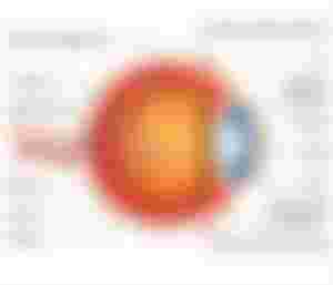
A groove is seen in the middle of the retina and near the blind spot. This is phobia. There are lots of conch cells but no rod cells. Much of our philosophy depends on it.
Blind spot (optic disk / blind spot)
This is the endpoint of the philosophy nerve. There are no photosensitive cells (rods and cones).
Optic nerve:
It is the second cranial nerve in humans. Through this the light sensation from the eyes reaches the brain
Thanks for Reading this. I hope it's helpful for us 💞 💕
Very informative article... thanks for sharing