Today a 34 year old male came to my office. He says that he has been having abdominal pain for a long time and he has also felt pain towards his pelvic area and back. This is what is known in medicine as referred pain. This is because this patient's gallbladder is presenting alterations.
The gallbladder is a hollow organ that is in charge of lodging a liquid produced in the liver called bile. This human viscera is located in a pit below the liver, it’s pear-shaped and the liquid stored in it is drained into the intestine through a duct called the common bile duct.

The gallbladder must contain only the biliary liquid to empty properly. After we consume food, the gallbladder physiologically contracts and drains its juices towards the intestine to intervene in the digestive process. But what happens when the gallbladder has obstacles in its interior?
In this case, I’ll only tell you what happens when the problem is that there are stones or as it is called in medical terms, kidney stones.
The gallbladder contracts involuntarily. In other words, it empties itself without our control, it contracts and expels the bile into the common bile duct. If there are stones inside it, it will continue to contract because that is its function, that’s when the pain appears in the patient. This happens because the gallbladder insists on emptying but the stone (or stones) inside it prevent it from doing so properly. The stones move, obstruct the exit orifice, that is, the common bile duct, and since the stone is larger than the normal diameter of the common bile duct, the stone cannot come out and the bile does not come out either, or it does so in very small quantities.
This persistence of the gallbladder to do its emptying and the constant impediment by the presence of the stone inside it make it inflamed. Of course all this is happening to the gallbladder of a human being. It should at least present mild to severe pain while it is doing its function.
Thanks to the presence inside the gallbladder's interior, the gallbladder remains with fluid inside to the point that it dilates too much. In order to counteract this great effort of the gallbladder, its walls become thicker and thus the pain increases in the patient. With time the pain becomes unbearable or becomes an emergency pathology due to the fact that the small stone or lithium insists on coming out through the orifice which is not large enough, sometimes becoming embedded and thus causing a surgical emergency called Acute Colecystitis.
It could also be the case that it’s a larger stone, which will also try to come out but because of its size it just can’t, causing an obstruction.
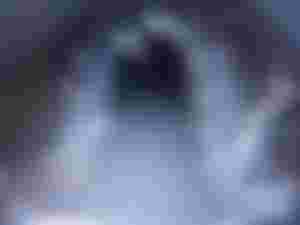
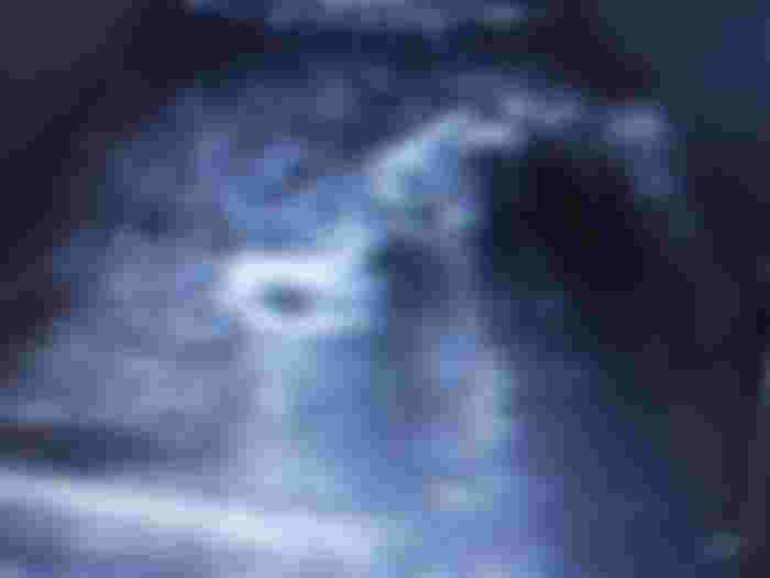
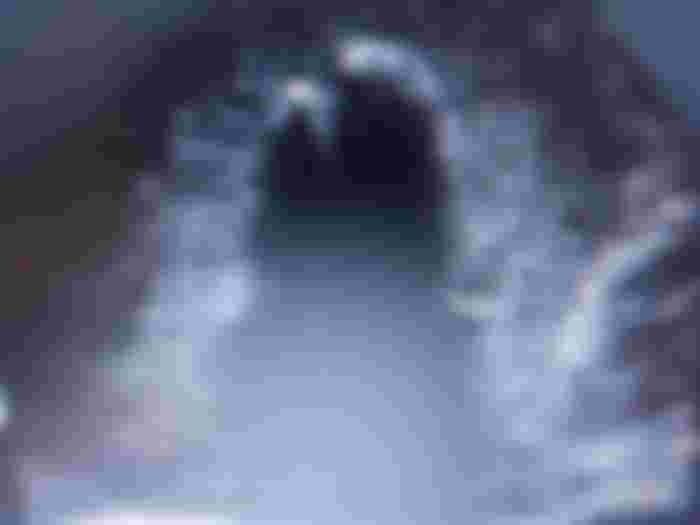
The patient I saw today in my office has so many stones in their gallbladder that the interior is not exactly hollow and black but presents this white linear surface and under that white line you can see the totally black shadow from the stones. This because the echo that lets me see is returned and does not penetrate these stones.
In this other picture there’s can some kind of convex image and inside or glued to it we have these stones, which stops the sound so the dark shadow is the only result that can be seen after the sound is lost in the depth.

This patient needs to go through a special diet until he can go to his doctor's appointment to have his gallbladder removed. Since the gallbladder is not fulfilling its function due to the impediment with the stones.
A simple diet is to avoid eating carbohydrates, red meat, white meat in small amounts, no chocolate, citrus, caffeine, condiments, fried foods, fats, etc… In this case, he should eat in small quantities and if possible 4 or 5 times a day or every three or four hours.
It’s necessary to remove the gallbladder as soon as possible because as long as these stones are inside the patient will always have pain. The diet is only to minimize the pain. It can’t be avoided, the discomfort will continue because the gallbladder contracts at will and we don’t know when the pain will be severe and medication is necessary in this case.
I hope this simple explanation can be of help to those who present this pathology.


ohh so that's what a gallbladder looks like under an ultrasound machine. it must take a lot of practice to be able to interpret the image