Tibia
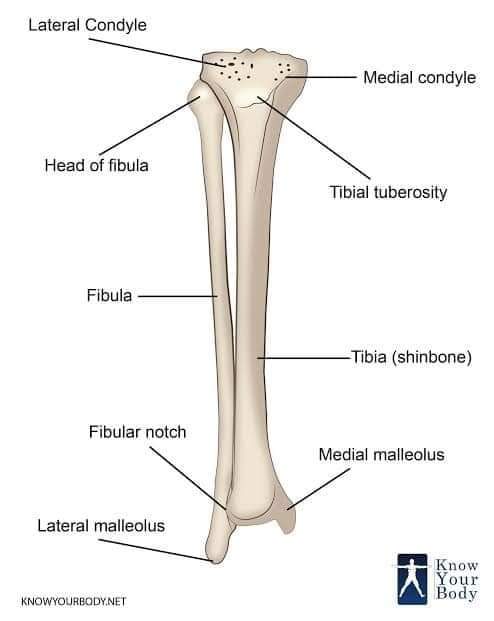
Tibia is a typical long bone. Cause, it has 2 epiphysis & 1 diaphysis. It transmits body weight from femur to foot. 🔥Has 3 ends: Upper end, Shaft & Lower end) ⭕ Upper end: has medial condyle, lateral condyle & tuberosity. ➡️ Medial condyle has medial meniscus (oval shaped). It's function is to prevent friction. It faces injury more. ➡️Lateral condyle has lateral meniscus (rounded). 🎉Meniscus is one type of fibro-cartilage) 🎉 Functions of meniscus: -Deepens the articular surface - transmits weight - absorbs shock -helps in gliding & angular movement 🎉 meniscus has a vascular structure which gets nutrition from Synovial fluid. ⭕ intercondylar area is situated between the two condyles. 🎉attachment from before backwards: 🔥#mnemonic- এমি একটি এলাকায় পলি এবং প্রমিকে প্রখর রোদে দেখেছে। এমি- Anterior horn of medial meniscus (এখানে 'এ' মানে anterior & 'মি' মানে medial) একটি- Anterior cruciate ligament. এলাকায়- Anterion horn of lateral meniscus. পলি- Posterior horn of lateral meniscus প্রমি- Posterior horn of medial meniscus. প্রখর- Posterios crutiate ligament. ⭕Tibial tuberosity: এটির Upper smooth part এ থাকে ligamentum patellae যেটি quardiceps femoris এর tendon থেকে এসছে। আর Lower rough part হলো subcutaneous, শুধুমাত্র infrapatellar bursa দিয়ে skin থেকে পৃথক থাকে। ⭕Knee joint এ intra-articular structure, tendon of popliteus, থাকে। ⭕Shaft: Shaft এর anterior border এর upper 2/3 কে #shin বলে। অ্যাথলেট বা প্লেয়ার রা খুব ভালো চিনবেন এই শিন। যেহেতু আপনারা #shin_guard ইউজ করেন। 🎉Medial surface- এটি পুরাটাই খালি একটা জায়গা। তবে, একদম উপরের দিকে ৩ টি muscle inserted হয়- -sartorius -gracilis -semitendinosus এদেরকে একসাথে #guy_ropes muscle বলা হয় কারণ এই ৩টি muslce thigh এর ভিন্ন ভিন্ন ৩টি compartment থেকে এসেছে। তাই nerve supply ও ভিন্ন ভিন্ন। 🎉Lateral surface: এখানে উপরের ২/৩ ভাগ জুড়ে rise করে Tibialis anterior. 🎉Nerve supply- Deep peroneal nerve. আর নিচের এক তৃতীয়াংশের সাথে related (medial to lateral): 🔥#mnemonics- আমার হাঁস একটি পঁচা ডিম পেরেছে। আমার- Tibialis anterior হাঁস-Extensor hallucis longus একটি-Anterior tibial vessels পঁচা-Deep perineal nerve ডিম-Extensor digitorum longus. পেরেছে-Peroneus tertius মজার না? 😂 হুম, এনাটমি বেশ মজার! যাই হোক, 🎉Posterior surface: একটি oblique soleal line দিয়ে দুই ভাগে ভাগ করা হয়। এই line এ থাকে soleus muscle.. Line এর উপরে থাকে Popliteus. এবং Line এর নিচে- ➡️Medially - Flexor digitorum longus. ➡️Laterally - Tibialis posterior muscle. এই দুটি মাসল কেই nerve supply দেয়- Tibial nerve. ⭕Lower end: One process (Medial malleolus) 5 surfaces (anterior, medial, posterior, lateral & inferior) 🎉️Medial malloelus এর lower border এ deltoid ligament of ankle joint attached থাকে । 🎉Lower end এর inferior surface, foot এর short bones গুলোর মধ্যে শুধুমাত্র Talus এর superior surface এর সাথে joint form করে। 🎉Tibia তে #subcutaneous অর্থাৎ বাইরে থেকে হাত দিয়ে স্পর্শ করে tibia এর যেইসব জায়গা অনুভব করা যায়- 1️⃣Rough area of tibial tuberosity. 2️⃣Shin of the tibia. 3️⃣Medial surface. 4️⃣Medial malleolus. (Total 4 nos.) 🎉️Tibia যেসব joints form করে- 1️⃣Knee joint- (Between two condyle of femur & two condyle of tibia) Type-Condylar variety of synovial joint. 2️⃣Superior tibio-fibular joint- (Between posterior surface of lateral condyle, fibular facet & head of fibula) Type- Plane type of synovial joint 3️⃣Middle tibio-fibular joint- (between two interoseous border of tibia & fibula) এই joint টির উপরে এবং নিচে দুটি gap থাকে। যার উপরের gap দিয়ে anterior tibial vessels গুলো anterior compartment এ ঢুকে & নিচের gap দিয়ে perforrating branch of peroneal artery আসে anterior compartment এ। Type- syndesmoisis type of fibrus joint. 4️⃣Inferior tibio-fibular joint- (between fibular notch of tibia & lower end of fibula) Type- Syndesmoisis type of fibrous joint. 5️⃣Ankle joint- (Between inferior surface of lower end of tibia & superior surface of talus) Type: Hing type of synovial joint. 🎉Growing end- upper end.. 💊Clinical: Tibia এর lower 1/3rd এ poor blood supply. তাই, non union of fracture common lower end এ।
No comments yet