Hip Bones
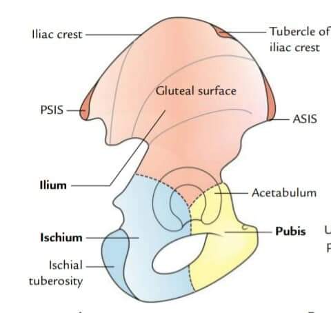
🔴There are two hip bones(Right& Left) The bone is a large irregular bone,articulates in front with the corresponding bone of the opposite side. Posteriorly, two hip bones articulate with the sacrum.
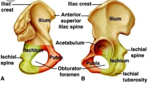
🔴️Parts:1) Ilium, 2)Ischium & 3)Pubis. 🔴️Side determination:- 1.The acetabulum directed laterally. 2.The flat,expanded ilium forms the upper part of the bone,that lies above the acetabulum. 3.The obturator foramen lies below the acetabulum. 🔘Ilium:- 🔶2 ends: 1)Upper end(Iliac crest) 2)Lower end-samller,fused with pubis & ischium in the acetabulum. Forms:upper 2/5th of acetabulum. 🔶3 borders:Anterior,Posterior &Medial. 🔶3 surfaces:Gluteal,Iliac fossa & Sacropelvic surface. 🔷Iliac crest:- 🔸2segment; Long Ventral segment(outer lip, inner lip,intermediate area); Short dorsal segment(Medial & Lateral slope). 🔶Ventral segment- 🔹Outer lip: ♦Attachment-Fascia lata(whole extent) ♦Origin:Tensor fascia lata(In front of tubercle) & Lattisimus dorsi(Behind the highest point of crest) 🔹Inner lip: A)Anterior 2/3rd- ♦Origin:Transversus abdominis ♦Attach:Fascia transcersalis &Fascia iliaca. B)Posterior 1/3rd- ♦origin:- Quadratus lumborum ♦Attach:-Thoracolumbar fascia. 🔶Dorsal segment: 1)Lateral slope:- origin- Gluteus maximus muscle.
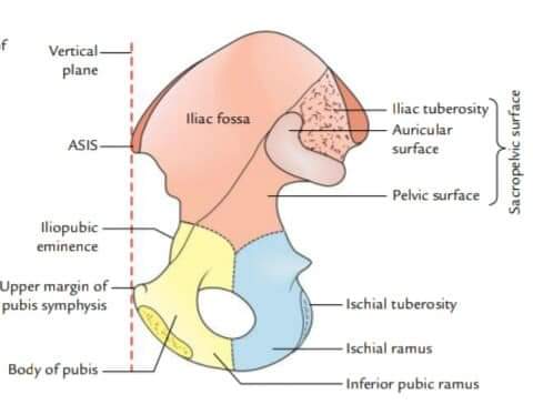
2)Medial slope:- Origin-Erector spine muscle. 🔹Anteriorsuperior iliac spine:- ♦Attachment-Lateral end of inguinal ligament. ♦Origin:Sartorius muscle. 🔴️Borders:- 1.Anterior border- ★Extend form ant. superior iliac spine to acetabulum. ♦Origin-Straight head of rectus femoris. ♦Lower part attchment-Iliofemoral ligament. 2.Posterior border- ★Extend from posterior sup.iliac spine to posterior border of ischium. ♦Attachment: Upper part-Sacro tuberous ligament 3.Medial border- ♦Inserted to middle arcute line:Psoas minor. Ventral sacro iliac ligament 🔴️Surfaces:- 🔘Gluteal surface: Divided by 3 lines- i)Posterior gluteal line; ii)Anterior gluteal line, iii)Inferior gluteal line. ♦Origin of mucle:- a)Gluteal maximus b)Gluteal medius c)Gluteal minimus d)Rectus femoris 🔘Iliac fossa:- ♦Origin-Iliacus muscle from upper 2/3rd. 🔘Sacro-pelvic surface:- 1)Iliac tuberosity- 🔹Attachment(Above to down): a)Interosseous sacroiliac ligament b)Dorsal sacroiliac ligament c)Iliolumber ligament. 2)Auricular surface:- 🔹Attachment -Ventral sacroiliac ligament(convex margin)
No comments yet